
3D CT Imaging
3D CT Imaging
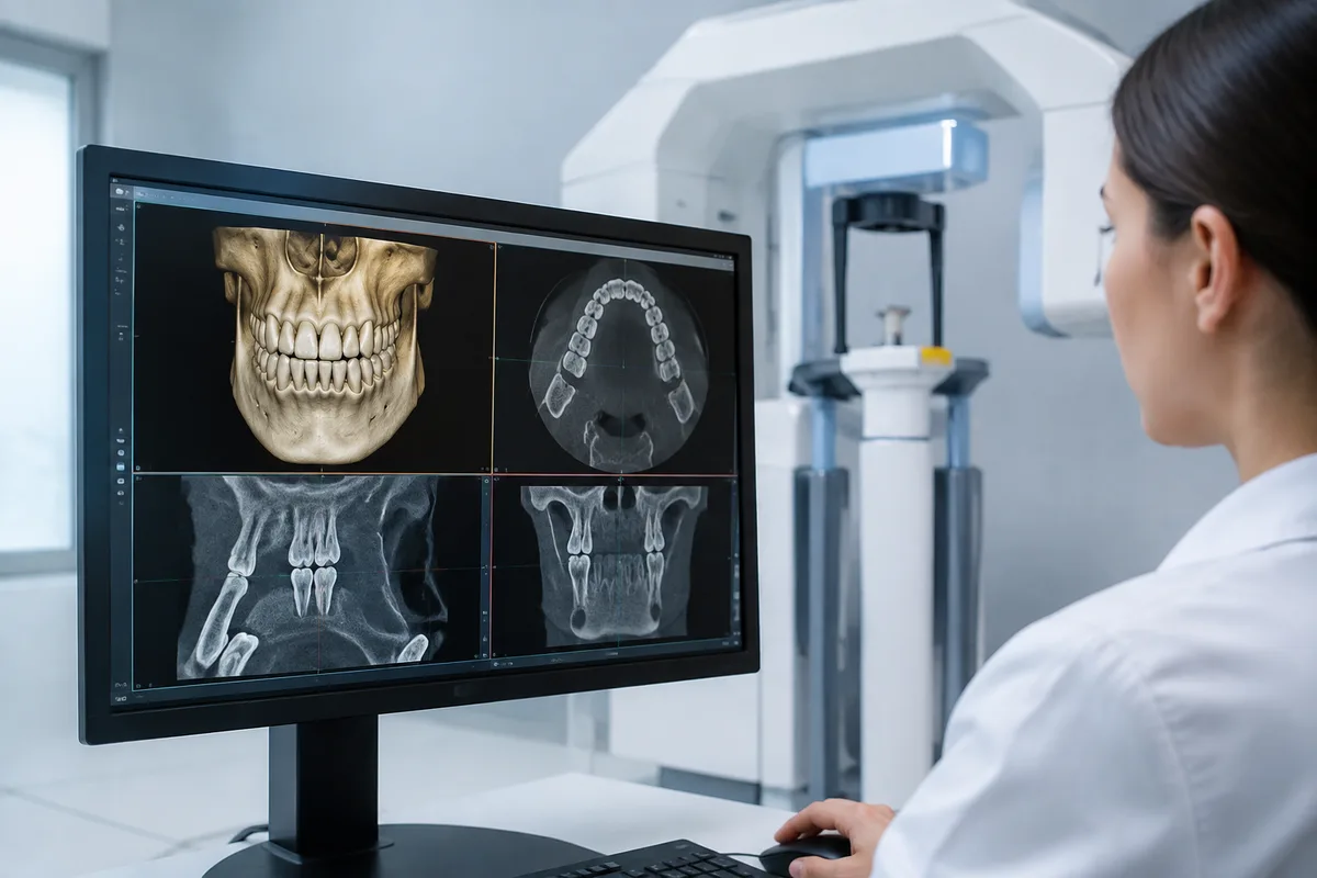
3D CT imaging is a single scan that captures teeth, soft tissue, nerve pathways, and bone density together, giving a complete picture of the jaw before surgery or complex treatment begins. At 1st Family Dental in Chicago, this imaging supports the planning that goes into implants, extractions, and orthodontic treatment, helping our team map out care with a clear view of what lies beneath the surface.
What 3D CT Imaging is
Cone beam computed tomography (CBCT), often called 3D CT imaging in dental settings, rotates a scanning arm around the head to capture a series of images that are combined into a three-dimensional model. Unlike a traditional two-dimensional x-ray, which flattens the mouth into a single plane, a CT scan shows depth, width, and height at once — the position of nerves, the density and shape of bone, the location of roots, and the relationship between structures that would otherwise overlap on a flat image.
This technology built on earlier panoramic and periapical x-rays by adding a dimension those images could not offer. Where a panoramic film gives a wide but flattened view of the jaw, a 3D scan lets a clinician rotate, slice, and measure the anatomy from any angle. That level of detail is particularly useful anywhere precision placement or careful surgical planning matters.
How we use it at 1st Family Dental
Our team draws on 3D CT imaging most often when planning implant and surgical care. Dr. John Hur, an Oral and Maxillofacial Surgeon whose training emphasizes CT-guided skeletal mapping and surgical protocols, uses this imaging to plan implant placement and corrective jaw surgery with attention to bone structure and nerve position. Dr. Najeeb Abufarha's training in digital treatment planning and 3D imaging also supports implant diagnosis, placement, and restoration from start to finish.
In orthodontics, Dr. Sameer Al-Obaidi and Dr. Budi Kusnoto are both trained in applying 3D imaging to diagnosis and treatment planning, which can help clarify root and bone positioning ahead of orthodontic movement. Across the practice, 3D CT imaging works alongside our other on-site technology — including Digital Panoramic X-Rays and Guided Implant Surgery — so that a case can be reviewed from multiple angles before any surgical step is taken. This kind of imaging is commonly paired with services such as dental implants, All-on-4 implant dentistry, Implant in a Day, bone grafting, and wisdom tooth removal, wherever a detailed view of bone and nerve anatomy helps guide the plan.
What it means for you
A single 3D scan can reduce the need for multiple separate x-rays, since it captures bone, soft tissue, and nerve pathways in one pass. For patients considering implants or oral surgery, this can mean a clearer conversation with your dentist about what a procedure involves, since the images make it easier to see and discuss the anatomy together. Because the scan shows precise measurements of bone density and available space, it also supports more accurate planning before any surgical guide or implant is placed. The scan itself is quick and non-invasive, requiring no injections or special preparation beyond what your dentist advises.
What to expect during your visit
Before the scan, you will be asked to remove any metal jewelry, glasses, or removable dental appliances that could interfere with the image. You will stand or sit in the scanning unit while the arm rotates around your head; you simply hold still and, in many cases, bite gently on a positioning guide. The scan itself typically takes well under a minute, though the full appointment — including positioning and any explanation of what the images show — usually takes just a few minutes longer.
Afterward, your dentist or specialist reviews the three-dimensional images with you, often on a chairside screen, pointing out the structures relevant to your treatment. From there, the images become part of the planning process for whatever comes next, whether that is implant placement, an extraction, or an orthodontic treatment plan.
Frequently asked questions
Is 3D CT imaging safe?
Dental CT imaging uses a low, targeted dose of radiation focused on the jaw and surrounding structures. It is a standard tool used by dentists and specialists for surgical and orthodontic planning, and your provider will only recommend it when the information it provides is useful for your specific treatment.
Is the scan uncomfortable?
No. The scan is painless and non-invasive. You simply remain still for a short period while the scanning arm moves around your head; there is no injection, no sound beyond a quiet whir, and no recovery time needed afterward.
How long does the scan take?
The scan itself is very brief, generally completed in under a minute. The full visit, including positioning and a review of the images with your dentist, is still a short appointment compared to many other dental procedures.
Who might need this type of imaging?
3D CT imaging is typically used when a dentist or specialist needs a detailed view of bone and nerve anatomy — for example, ahead of implant placement, certain extractions, jaw surgery, or orthodontic treatment planning. Your dentist will let you know if your treatment calls for this type of scan.
If you have questions about 3D CT imaging or want to know whether it is part of your treatment plan, reach out to 1st Family Dental — we are happy to talk it through with you.